
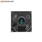
Separated Endoscope Camera Module
Your Professional Camera Module Manufacturer
Guangzhou Sincere Information Technology Ltd. is a professional and high-tech leading company in integrated optical device manufacturer and optical imaging system solution provider since 1992's foundation. We specialize in the production of various camera modules to help you create highly customized camera module solutions, including 0.3mp to 108mp MIPI camera modules and USB camera modules as well as endoscope camera modules with a diameter of 0.9mm ~8.0mm.
Quality Assurance
All our camera modules must be inspected by professional QC, and the products are inspected in strict accordance with national standards before shipment. And the entire process is strictly implemented in accordance with the ISO9001 quality system.
Advanced Equipment
Professional AA (Active Alignment) equipment manufacturing, COB 100 level dust-free workshop.
Professional Technical Team
We have been manufacturing camera modules for over 30 years. And we have top professional R&D talents, management talents and sales elites with rich experience.
Good Service
We provide 1-year replacement and 10-year warranty services. Additionally, we can provide training on how to use the camera module.
Reasonable Price
We offer competitive price to achieve win-win.
Wide Range Of Applications
Specializing in machine vision, we provide cutting-edge camera modules that support a wide range of applications in various industries, meeting the needs of everything from agriculture to healthcare, industrial automation to retail, SINCEREFIRST is the trusted partner that fuels these applications with unparalleled machine vision excellence.


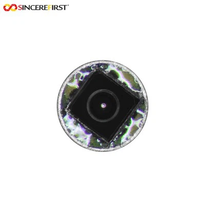


The Separated Endoscope Camera Module is a type of module that separates the camera part from the other components of the endoscope. It usually consists of a miniaturized camera and connection cables, etc. The camera can be installed at the tip of the endoscope to capture images of observation areas such as inside the body or inside the equipment. The image signals are transmitted to external image processing devices or displays through the cables. This module design is convenient for flexible installation and use, can meet the needs of some special application scenarios, such as different types of endoscope examinations and surgeries in the medical field, which can improve the applicability and operational convenience of the equipment, and is also conducive to cleaning and disinfection, reducing the risk of cross-infection. At the same time, it can also be used for high-definition imaging detection in narrow spaces in industrial inspection fields.
Advantages Of Separated Endoscope Camera Module
Flexible and Portable Operation
The separated design enables a reasonable distribution of the endoscope's weight. In addition, the camera module can be independently adjusted, which is convenient for detecting in complex spaces.
Convenient Disinfection and Maintenance
Some medical use separated endoscopes can separate the part in contact with the human body from the control part. This can shorten the pollution path, make the disinfection process more convenient.
Excellent Imaging Quality
The separated endoscope camera module can be equipped with a high - performance imaging system, can provide high - definition and distortion - free dynamic images.
Realization of Multiple Functions
The separated design facilitates the upgrading and expansion of functions, the camera module can be replaced or matched with different light sources to adapt to different detection environments and needs.
Reduced Costs
The replaceable camera module can reduce the overall maintenance cost, and when a problem occurs, only the damaged part needs to be replaced, without replacing the entire device.
Ultra-Fine Lens Diameter Design
Through the separate design of the lens and the circuit board, the diameter of the lens can be reduced to about 1mm, enabling it to fit into the narrowest gaps.
Types of Separated Endoscope Camera Module

Bare Separated Endoscope Camera Module
A basic type without additional protective casings, consisting of a miniaturized camera, essential sensors, and connection cables.
Steel Shell Separated Endoscope Camera Module
Equipped with a rigid steel sleeve around the lens and cable, this type enhances durability and resistance to mechanical stress.


HD Separated Endoscope Camera Module
Emphasizes high-definition imaging performance, integrating advanced sensors and image processing technology to deliver clear, distortion-free visuals.
LED Separated Endoscope Camera Module
Incorporates miniaturized LED lights directly into the camera tip, eliminating the need for external lighting sources. The separated design places the LED control circuit outside the lens, ensuring the lens remains ultra-fine.


Ultra-Wide-Angle Separated Endoscope Camera Module
Equipped with an ultra-wide-angle lens to capture a broader range of the target area in a single shot. The separated design places the lens control components externally, ensuring the lens remains compact while achieving wide-angle coverage.
Ultra-Fine Diameter Separated Endoscope Camera Module
Specialized in minimizing the lens diameter (as small as 0.9mm) through optimized separation of the lens and circuit board.

Application of Separated Endoscope Camera Module

Medical Gastrointestinal Examinations
The ultra-fine lens enters the gastrointestinal tract smoothly, capturing high-definition images. Its detachable, disinfectable camera reduces cross-infection risks, with flexible angle adjustment aiding in detecting small lesions.

Minimally Invasive Surgeries
Lightweight and easy to maneuver, it provides real-time, clear surgical site images. Replaceable modules minimize downtime during procedures, assisting precise operations.

Industrial Pipeline Inspection
Ultra-fine lenses access narrow pipeline gaps to check for corrosion, cracks, or blockages. Flexible camera adjustment and high-definition imaging ensure comprehensive defect detection.

Electronic Component Quality Checks
Fits into micro-component gaps to inspect internal structures. High-quality imaging identifies soldering issues, dust, or damage, ensuring electronic product quality.

Respiratory Tract Diagnostics
Ultra-fine lenses cause minimal patient discomfort. Disinfectable cameras meet medical standards, with clear images aiding in observing respiratory tract abnormalities.

Large Machinery Inspections
Flexible operation and wide-angle lenses (when applicable) access narrow spaces in machinery. Detects wear or looseness in components, reducing failure risks and maintenance costs.
Process of Separated Endoscope Camera Module
1. Design Phase
Requirement Analysis and Specification Definition: Based on the specific application scenarios of customers, determine the core parameters of the module, including lens diameter, resolution, interface type, waterproof level, imaging frame rate, etc.
Hardware and Structural Design: Design the PCB layout to match the camera sensor, lens module, LED fill light and control chip; simultaneously design the shell structure to ensure the separation of the lens and the circuit part, taking into account portability and protection.
Prototype Development and Verification: Produce prototypes, test imaging effects, interface compatibility, temperature resistance, etc., optimize design defects and then finalize the design.
2. Production Phase
Material Procurement and Quality Inspection: Purchase qualified raw materials (lenses, sensors, PCB boards, connectors, shells, connection cables, etc.), and screen qualified materials through appearance inspection and performance sampling inspection.
PCB Board Mounting (SMT): Solder electronic components such as control chips and sensors to the PCB board through surface mount technology to ensure firm solder joints and unobstructed circuits.
Preliminary Testing and Solder Repair: Conduct power-on tests on the PCB board after mounting, check whether the circuit is short-circuited and whether the components work normally, and repair defective solder joints.
AA Process (Active Alignment Process):
This is a core step to improve imaging accuracy. Precisely align the lens module with the sensor through automated equipment:
Fix the lens holder at the preset position of the PCB board, power on the sensor and output real-time images;
The equipment captures the sensor imaging through the optical system and analyzes parameters such as clarity and distortion rate;
Drive the robotic arm to fine-tune the X/Y axis position and pitch angle of the lens until the image reaches the optimal state;
Fix the lens and the holder with UV glue, and complete the alignment after ultraviolet curing to ensure that the module imaging is free of offset and blur.
Module Assembly: Assemble the lens components that have completed the AA process with the shell and connection cables, integrate the LED fill light (if applicable), ensure the structure is stable, and the lens can flexibly adjust the angle to meet the separated design requirements.
Function and Performance Testing:
Connect to external image processing devices or displays to test the stability of signal transmission, the clarity of image transmission, and whether the frame rate meets the standards;
Detect waterproof performance, flexibility of lens adjustment and brightness of fill light (if applicable);
Screen out defective products with abnormal imaging, signal transmission failures, etc.
Cleaning and Appearance Inspection: Perform dust removal and cleaning on qualified modules, especially the detachable camera parts, and check whether the shell has scratches and whether the interface is intact.
3. Packaging and Shipping
Packaging: Put the module into an anti-static bag separately, then put it into a packaging box printed with product information (model, parameters, quality inspection mark), with built-in buffer materials to prevent damage during transportation; attach instructions, warranty cards and other accessories, and mark the detachable parts for proper handling.
Warehousing: Store the packaged products in a constant temperature and humidity warehouse in batches, and keep good inventory records.
Shipping: Sort products according to order information, ship through logistics channels, and provide logistics tracking information simultaneously to ensure that the products are safely delivered to customers.
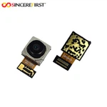
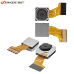
Components of Separated Endoscope Camera Module
Miniaturized Camera Unit
Lens: Ultra-fine lens (as small as 1mm in diameter) designed for accessing narrow spaces, with options for wide-angle or high-definition configurations to adapt to different observation needs.
Image Sensor: Typically a CMOS or CCD sensor that converts optical signals into electrical signals, responsible for capturing high-definition, distortion-free images.
Protective Casing: Optional steel sleeve or medical-grade plastic housing (for the camera tip) to enhance durability, resist corrosion, or meet sterility requirements (especially in medical applications).
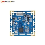
Imaging Control Module
Circuit Board (PCB): Separated from the camera unit, it integrates chips for image processing, signal amplification, and data conversion, ensuring stable image output and adjusting parameters like brightness or contrast.
Control Chip: Manages sensor operations, coordinates with external devices, and optimizes image quality in real time.
Lighting System
LEDs or Fiber-Optic Illuminators: Built into the camera tip (LEDs) or connected via separate fibers (fiber-optic) to provide uniform, high-intensity light for clear imaging in dark environments (e.g., internal body cavities or industrial pipelines).
Light Control Circuit: Separated from the camera unit to avoid increasing the lens diameter, regulating light intensity based on the observation scenario.
Connection Components
Transmission Cables: Flexible, thin cables (e.g., coaxial or USB cables) that transmit image signals from the camera unit to external devices (displays or processors), ensuring low signal loss even over extended lengths.
Connectors: Detachable interfaces (e.g., medical-grade or industrial-standard plugs) that enable easy separation of the camera unit from other components for disinfection, replacement, or maintenance.
Mechanical Adjustment Parts
Adjustable Brackets: Allow precise positioning or angle adjustment of the camera unit, facilitating flexible operation in complex spaces (e.g., bending around corners in pipelines).
Fixing Structures: Secure the lens and sensor during the AA (Active Alignment) process and maintain stability during use, preventing misalignment that could degrade image quality.
Protective and Auxiliary Components
Waterproof Seals: Applied to the camera tip and cable junctions to ensure water resistance (critical for medical or industrial wet environments).
Anti-Static Coatings: Protect sensitive electronic components from static damage during handling or operation.
How To Cooperate With Us?
Demand Analysis
Communicate requirements with customers
Design Scheme
Design solutions that meet customer needs
Establish Cooperation
Provide camera module drawings and establish cooperation
Make Samples
Camera module proofing according to the design plan
Camera Module Test
Send out samples, and customers will test
Mass Production
After the samples pass the customer's test, mass production begins
Certifications
RoHS, REACH, ISO, CE, FCC
")
FAQ
Q: What is an endoscope camera used for?
A: Endoscopy is the use of cameras to relay images from inside the body. The device used, known as an endoscope, is a long, thin, and flexible tube that has a camera at one end as well as a light source. An endoscopy can be used to diagnose a wide range of conditions, and the results are interpreted by a specialist.
Q: What is the difference between a borescope and an endoscope camera?
A: Endoscopes are more flexible, they cost more and they are used most commonly in medicine and veterinary care. Borescopes are less flexible, they cost less, they offer a better field of view and they are used much more widely across many different industries.
Q: How does an endoscope device work?
A: Fiberoptic endoscopic devices consist of flexible tubing that contains a series of lighted mirror lenses and optic fibers. These instruments transmit light around corners, twists, and bends, allowing direct visualization of body systems not easily visualized by other means.
Q: What makes a separated endoscope camera module "separated"?
A: It splits the camera unit (lens, sensor) from the control circuit, cables, and other components, enabling flexible operation and easy disinfection.
Q: What’s the typical lens diameter of such modules?
A: As small as 0.9mm, allowing access to ultra-narrow spaces like tiny pipelines or delicate body cavities.
Q: What organs can a doctor see during endoscopy?
A: An endoscopy procedure involves inserting a long, flexible tube called an endoscope down your throat and into your esophagus. A tiny camera on the end of the endoscope allows views of your esophagus, stomach and the beginning of your small intestine, called the duodenum.
Q: Will an endoscopy show liver problems?
A: An endoscopy can detect many types of liver diseases as well, such as cirrhosis, esophageal varices, and portal hypertension. Liver diseases can include: Hepatitis.
Q: How to ensure sterility for medical use?
A: The detachable camera unit can be thoroughly disinfected, and some use medical-grade materials to lower infection risks.
Q: Why is the AA Process important?
A: It precisely aligns the lens and sensor via automation, ensuring clear, distortion-free imaging.
Q: Are they suitable for both medical and industrial use?
A: Yes. Medical versions focus on disinfection and precision; industrial ones excel in narrow-space inspections.